Surface-constrained Volume Registration (SVReg)
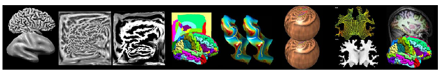
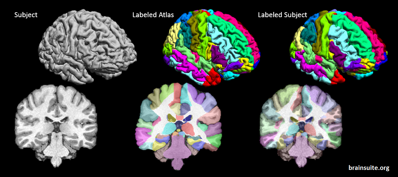
SVReg stands for Surface-constrained Volume Registration. SVReg is an algorithm and a software tool for co-registration of human-brain MR images. It uses anatomical information from both the surface and volume of the brain images for accurate automated co-registration which allows consistent surface and volume mapping to a labeled atlas. Brain labeling is a necessary preprocessing step for several studies looking at group comparisons and regional brain analysis.
Registration Model
SVReg utilizes a series of multi-step registration and refinement processes based on several consideration of useful morphological features and known variation of human brain anatomy. It has the ability to analyze both normal and abnormal brains of subjects starting at least 4 years old, even working well with lesion brains.
Our Atlases
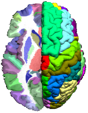
SVReg has been customized for registration of individual subjects to the atlas templates provided within BrainSuite’s installation package. Conversely, the provided atlas templates were created to cater to SVReg’s registration model for optimal results. Within the GUI interface, users may choose from three atlases that have been preprocessed and segmented by expert neuroanatomists.
Additional atlases are available for download using the links below, and can also be used with BrainSuite. We also include utilities with these atlases to facilitate their use with Freesurfer and FSL. To use these atlases with BrainSuite, download the atlas that you are interested in using the links below, unzip and move the folder to BrainSuite/svreg folder in your installation.
 The BCI-DNI_brain atlas, USCBrain Atlas, and USCLobes Atlas are each licensed under a Creative Commons Attribution-NonCommercial-ShareAlike 4.0 International License
When using theses atlases in your published work, please cite the following reference paper:
Joshi AA, Choi S, Chong M, Sonkar G, Gonzalez-Martinez J, Nair D, Wisnowski JL, Haldar JP, Shattuck DW, Damasio H, Leahy RM (2022) A Hybrid High-Resolution Anatomical MRI Atlas with Sub-parcellation of Cortical Gyri using Resting fMRI. Journal of Neuroscience Methods 374:109566.
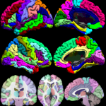
BCI-DNI Brain Atlas
 The BCI-DNI_brain atlas is a high-resolution atlas that was created with our collaborators at the Brain and Creativity Institute’s Dornsife Neuroscience Imaging Center at the University of Southern California.
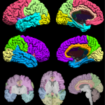
USCBrain Atlas
The USCBrain atlas uses the same single subject MRI data as BCI-DNI atlas but provides a finer subparcellation of cortical gyri based on functional parcellation of each gyrus. The USCBrain Atlas is now included in the BrainSuite21a distribution and can also be downloaded separately.
BrainSuiteAtlas1
 The BrainSuiteAtlas1 is based on the Colin27 atlas and is a stereotaxic average of 27 scans of an individual which is widely used by the neuroscience community.
USCLobes Atlas
 The USCLobes atlas segments the brain into larger regions than those provided by the default BrainSuite atlases. The detailed description of the atlas can be found here.
Deformation map from atlas to subject
By default SVReg will output:
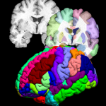
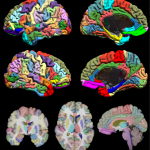
- Labeled brain surfaces and volumes into segmentations of ~90 ROIs
- Computation of GM, WM, CSF, total volume, average cortical thickness, and surface area of each ROI
- Vertex-wise cortical thickness mapped to atlas surfaces
- Delineation of up to 76 sulci on the mid-cortical surface
Optional Customization
Users may choose to run SVReg through the GUI or through command-line for batch processing and customization.
Users have the option to choose from a number of computational flags or designate manual inputs for highly controlled registration including manual masks, sulcal inputs, and atlases. SVReg can additionally be run in a one step process or be run piecemeal by executing individual modules.
Additional Tools
Additional tools are provided for users who would like to further analyze SVReg.
Our data processing package includes stand-alone functions to read and write SVReg outputs and main scripts to perform the following functions:
- ROI-wise group comparison
- Vertex-wise group comparison
- Surface smoothing
- Regenerating statistics after manual corrections
- Creating custom atlas
- Transforming data to atlas space
- Edit ROIs on surfaces and transfer edits to volume labels
Additionally we provide tools for: