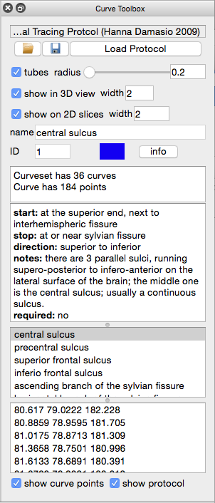
Sulcal and gyral landmarks on the human cerebral cortex are the primary means by which we identify cortical anatomy in MRI images. Curves representing sulcal fundi and gyral crowns can be used to constrain cortical surface coregistration in intersubject analyses for group studies of morphometric variability and functional activation. We have developed an interactive tool for performing landmark delineation (Shattuck et al., 2009a), which is built into our BrainSuite interface. For a given landmark, the user first clicks on a seed point. The user then selects a second point on the path of the landmark, e.g., at the opposite end of a sulcus. The algorithm applies Dykstra’s shortest path algorithm, which operates on the edges of the surface mesh. To track sulcal fundi or gyral crowns, our approach modulates the length of the edge by a measure of curvature at the vertices of each edge. The user may select additional points along the curve to define more complicated landmarks (e.g., an interrupted sulcus). The GUI for curve delineation also provides a facility for loading a detailed protocol specification that will guide a user through the process of delineating a cortex model, such as the widely used protocol developed by Dr. Sowell at UCLA or the 26 curve protocol developed by Dr. Hanna Damasio at USC as described in our recent paper (Pantazis et al., 2010).

Curve Tracing in BrainSuite
BrainSuite includes tools for delineating curves that identify sulcal or other cortical landmarks.
To use these tools with a cortical surface model derived from a 3D MRI volume, do the following:
- Start BrainSuite
- Load a volume file
- Load a surface model
- These can both be loaded by dragging-and-dropping the files onto BrainSuite.
- Open the Curve Toolbox (Tools→ Curve Tool from the main menu, or right click on tool bar→ Curve Tool Sidebar, or press c key)
- Select a curve to trace from the Curve List
- Select points on the path of the structure you want to delineate.
- You can select points in either the surface or the volume.
- Shift + Left click lets you continue an existing curve.
- Alt + Left click selects the previous curve.
- The pending curve is drawn in white on the surface and volume views.
Important options:
- Change the color by clicking on the color box; this will update in both the volume and the curve drawings.
- By default, curves are simultaneously displayed in the volume view. To disable this, uncheck the “draw curves in volume view” box. You can also adjust the thickness of the curves in the volume view by changing the “line size” value.
- To turn off synchronization between the the surface and volume view, uncheck the “track position in volume” box.
Available Protocols
The following BrainSuite cortical delineation protocol files are available:
BrainSuite delineation protocol developed by Hanna Damasio, MD. This protocol is described in our NeuroImage paper (Pantazis et al., 2010) and is included with the BrainSuite11a release.
Protocols suitable for surfaces produced with the MNI tools:
Cortical delineation protocol developed by Dr. Elizabeth Sowell
Reduced version of the above protocol This is the protocol that was used in our J Neuroscience Methods paper (Shattuck et al., 2009).